Veterinary Thermograpy
The use of surface heat patterns to assess health is nothing new: “In whatever part of the body excess of heat or cold is felt, the disease is there to be discovered”, Hippocrates claimed in around 400 BC. He would spread thin layers of mud on his patients and then watch the drying pattern. You could say this was the first ‘thermogram’.
Although the military successfully used thermal imaging in the 1940s and 50s, these cameras were unsuitable for veterinary (animal) use and it was not until the 70s and 80s, when the cameras became slightly more portable, that the technology started to be used to look at animals. However, the resolution was poor and printers could only produce a black and white image.
In 1990, a new device known as a focal plane array was released from the military into the commercial marketplace. This was a significant improvement and the result was an increase in image quality, but it was only after 9/11, when the US government targeted thermal surveillance, that finally a truly portable camera was developed and the use ofthermography to assess animals really began. Today, horses and dogs are probably the most commonly scanned but the technology can be used for any warm-blooded animal.

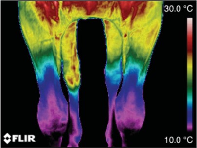
Figure 1. Cranial view, proximal fore limbs

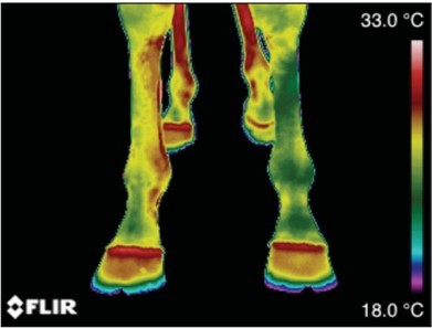
Figure 2. Dorsal view, distal fore limbs
Thermal imaging (thermography) does not use any radiation and is therefore perfectly safe for the animal and the handler. Extensive research in human and equine fields has demonstrated that many injuries and physical conditions can be accurately detected using thermography before any physical signs and symptoms are apparent.
Thermal imaging shows the animal’s physiological state by graphically mapping skin surface temperature in response to changes in blood flow, providing an objective view of the subjective feeling of pain. In healthy animals, the thermal pattern on the skin is largely symmetric; this is because skin blood flow is controlled by the sympathetic nervous system. Therefore, veterinary thermography can be an effective method for highlighting the location of problem areas, which then may require further investigation.
Thermal imaging is a complex and exacting science and requires a technician who is trained not only in animal physiology and anatomy but also infrared science and camera hardware and software. Currently there are no regulations, so unfortunately anyone can buy a camera and call themselves a veterinary thermographer, but fortunately there are ISO standards and American Academy of Thermology (AAT) Veterinary Guidelines for Infrared Thermography.
Anyone looking to employ the services of a veterinary thermographer should ensure that the thermographer has been well trained according to ISO standard 18436-7 and follows ISO standard 18434-1 and the AAT Veterinary Guidelines for Infrared Thermography, that they obtain pre- and post-exercise scans and can produce an industry-standard report.

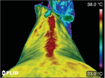
Figure 3. Thoracic/lumbar back

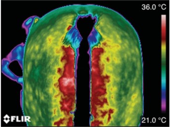
Figure 4. Caudal hindquarters
In the equine application, a well-trained and experienced thermographer will take a full body scan both before and after exercise adhering to strict protocols. The thermographer’s report will contain 100-120 images with an interpretation of the thermal patterns. This can give the owner, vet or other equine professional further information that they might not otherwise know, including ‘areas of interest’ and how the horse is weight bearing. Thermography is physiological imaging: it shows what is happening, as opposed to anatomical imaging such as radiographs and ultrasound, which show what has happened.
